Structuring a Retinal Imaging Workflow When OCT and Fundus Imaging Are Separate
A practical framework for reviewing friction points between OCT, fundus imaging and possible angiography, with the HOCT-1/1F presented as a combined-equipment option.
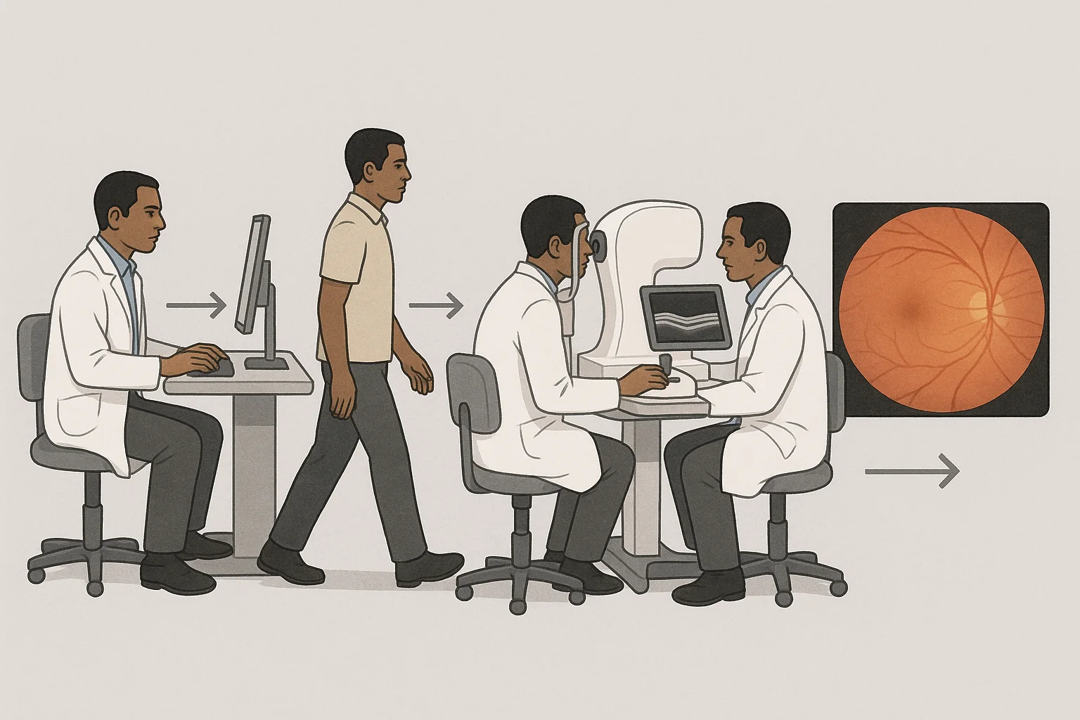
The professional answer starts with workflow mapping, not with an immediate equipment decision. When OCT and fundus imaging are performed separately, the practice should make every step visible and review the transitions between systems.
1. Map the current workflow
Describe the pathway as it operates today: patient arrival, OCT acquisition, fundus imaging, then consultation or image review. The purpose is to identify where the patient, operator or information moves from one step to the next.
Questions to document
- Which examination comes first?
- Does the patient move to another room or operator?
- Are the images reviewed within one coordinated work sequence?
- At what point might angiography be considered?
2. Identify workflow friction
With separate systems, the key issues are organisational. They include examination sequencing, coordination between workstations and continuity during image review. These questions should be assessed with the teams who use the workflow in practice.
A retinal imaging workflow in an ophthalmology practice should be assessed through the steps actually followed, not only through the list of available devices.
3. Consider combined equipment
The official “HOCT-1/1F” brochure covers the HOCT-1 and HOCT-1F models. Huvitz presents the HOCT-1F as equipment combining OCT and fundus imaging.
The more useful question is not only “Which device should we choose?” but also “Which steps are we trying to bring closer together or simplify?” The answer depends on the current pathway and on how the practice allocates examinations.
4. Keep other examinations clearly defined
Angiography may be treated as an optional part of the pathway according to the practice protocol. It should not be presented here as a function of the HOCT-1F, because the available evidence establishes only the combination of OCT and fundus imaging.
The same distinction applies to complementary equipment. Huvitz lists OCTavius in its ophthalmology and optometry offering. The HVF-100 is listed in the perimeter category of Huvitz’s diagnostic range for ophthalmology and optometry. These references can be recorded separately when the full diagnostic pathway is reviewed.
A simple decision framework for the practice
Before changing the workflow
- Describe the current pathway without removing operational detail.
- Separate combined examinations from examinations that remain organised independently.
- List transitions between workstations, rooms and operators.
- Compare the current organisation with the intended workflow.
- Record the selected approach in the practice’s internal protocol.
Share this article
Professional FAQ
What should a practice review first when OCT and fundus imaging are performed separately?
Start by describing the actual pathway: the order of examinations, movement between systems, patient flow and the way results are reviewed. This makes the practice-specific friction points visible.
Does the HOCT-1F combine OCT and fundus imaging?
Yes. Huvitz’s official brochure presents the HOCT-1F as equipment combining OCT and fundus imaging.
Is OCTavius part of Huvitz’s ophthalmology offering?
Yes. Huvitz lists OCTavius within its offering for ophthalmology and optometry.
Is the HVF-100 a retinal imaging device?
Huvitz’s official website places the HVF-100 in the perimeter category of its diagnostic product range for ophthalmology and optometry. It should therefore be considered separately when mapping the wider diagnostic workflow.